John Manak

Professor
Biomedical Sciences Program Director
Biography

Research summary

Genomics, genetics, neurobiology

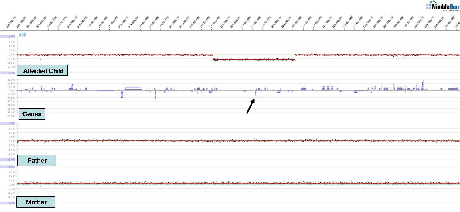
Work in my laboratory focuses on understanding human genetic disorders by first identifying gene mutations associated with those disorders and then modeling the disorder in relevant organisms. We have identified novel genes and chromosomal structural variants (see figure) associated with a variety of congenital anomalies including spina bifida, branchio-oto-renal syndrome, renal agenesis, and cleft lip and/or palate. These studies have led to publications in Hum Mol Genet, Hum Genet, Genetics (invited and highlighted articles), and Am J Hum Genet amongst others. Of particular interest is a new clefting and craniofacial patterning gene we identified, ISM1. We have found that knockdown of ISM1 in frogs inhibits migration of cranial neural crest cells, the key cells that make up the structure of the face, thus leading to severe craniofacial anomalies including clefts. We have also studied the fly homolog of the C-MYB proto-oncogene (Dm-Myb) where we discovered a new function for Myb in separating and stabilizing chromatin neighborhoods associated with topologically associating domains (TADs); loss of Dm-Myb results in loss of H3K27me3 repressive domains, leading to robust derepression of large numbers of genes in the genome. Interestingly, Myb is the primary DNA-binding factor that binds to TAD chromatin loop anchors in flies and may also be one of the key anchor proteins working alongside CTCF in humans. These studies were published in Nature, Nature Cell Biol, PNAS, and Cell Reports amongst others.   

A major focus of the laboratory is understanding the genetic basis of epilepsy, using a fruit fly model of an epilepsy-ataxia syndrome. We have shown that fly PRICKLE mutants exhibit a remarkably similar syndrome to humans carrying PRICKLE mutations (early-onset, progressive, myoclonic/tonic-clonic seizures as well as ataxia) and have identified enhanced axonal transport defects as one cause of the seizure phenotype. Recently, we have shown that activation of the glial innate immune response (IIR) leads to increased neuronal cell death which is responsible for exacerbating the seizure phenotype and that increases in neuronal oxidative stress ultimately lead to activation of the IIR. We thus provide the first direct genetic evidence that oxidative stress and the glial IIR can lead to the progression of epilepsy. These studies were published in PNAS, Am J Hum Genet, PLoS Genetics, Ann Clin Transl Neurol, and Cell Reports amongst others. Current studies are aimed at determining whether we can repurpose well-tolerated antioxidant and anti-inflammatory drugs as a new class of anti-epileptic drugs (AEDs). We are also exploring how the glia are activated in our seizure mutant; for example, are damage-associated molecular patterns (DAMPs) the culprit, and which ones? Additionally, we are exploring how the glial IIR is able to promote neuronal cell death, thus exacerbating the seizure phenotype.

Project 1

One of the primary areas of research in my laboratory is elucidating the genetic basis of neurological disorders, including epilepsy and Alzheimer’s Disease.  We have found that mutations in the prickle gene cause seizure disorders in fruit flies, similar to those observed in mice and humans with orthologous Prickle mutations (Tao, Manak, Sowers et alAJHG, 2011), and we are now using the fly model to perform both directed and unbiased genetic screens to identify other components in the seizure pathway, as well as elucidate the mechanism responsible for the disease phenotype. We have shown that mutations affecting different isoforms of the fly prickle gene have opposite behavioral phenotypes; remarkably, mutation of the pksple isoform makes flies susceptible to seizures, whereas mutation of the pkpk isoform actually makes the flies less susceptible to seizure activity.  This suggests that tipping the balance of prickle isoforms in one direction can drive the seizure phenotype, and consistent with this hypothesis we have been able to create an epileptic fly simply by increasing the pkpk isoform exclusively in neurons and muscles (mimicking the imbalance we see in the pksple mutants; Ehaideb et alPNAS, 2014).  We have analyzed the electrophysiology of the prickle mutants in collaboration with Chun-Fang Wu's lab and have found that the pksple flies have a reduced seizure threshold compared to control flies.  Additionally, we have identified the cellular process that is altered in the pksple mutants.  In a recently published study (Ehaideb et alPNAS, 2014), we show that anterograde vesicle transport in neurons is enhanced in the pksple seizure-prone mutants, and that we can fully suppress the seizure phenotype by reducing the dose of either of two Kinesin subunits, which make up the motor responsible for anterograde transport.  These data reveal a new pathway in the pathophysiology of seizure disorders, and have also provided several intriguing connections to neurodegeneration.  Since roughly one third of epilepsy patients have adverse effects from the drugs that are currently available, and since some of the more popular drugs have been associated with birth defects, there is a great need to develop safer and more effective anti-epileptic medications. We are thus using the pksple flies to screen novel anti-epileptic drugs; our pipeline from fly to mouse to human will allow us to move from one system to the other as we begin to identify molecules that promote anti-seizure activity. Although not described here, additional work in my laboratory is focused on using fruit fly genetics to functionally validate human gene mutations associated with neurodevelopmental disorders such as ID (intellectual disability) and ASD (autism spectrum disorders).

Project 2

Over the last several years, it has been demonstrated that genomic rearrangements play a role in human disease, which has in turn created new opportunities for finding the specific genes involved in the disease process. Genomic rearrangements can arise when interspersed repeat elements, lying in tandem, facilitate submicroscopic deletion (or duplication) events. Any gene caught in a genomic rearrangement could be identified by data demonstrating a change in that gene’s dosage. Tiling microarrays have been used to identify such changes using a procedure termed array-based Comparative Genomic Hybridization (aCGH). aCGH relies on competitively hybridizing to the tiling microarray a fluorescently labeled reference genomic DNA sample with a fluorescently labeled experimental sample from an individual afflicted with a disease. By comparing hybridization intensities of the reference and the experimental samples, it can be determined whether amplifications or deletions have occurred in the genomic region of interest. These changes are referred to as DNA copy number changes, or Copy Number Variants (CNVs). Importantly, CNVs are now considered common causes of human disease. aCGH has been used successfully to identify CNVs associated with a number of human diseases (for example, Kallioniemi, 2008; Walsh T. et al, 2008, Sharp et al, 2008, Ballif et al, 2007, Lenz et al, 2008). In collaboration with Drs. Alex Bassuk, Jeff Murray, Tom Wassink, Richard Smith and Patrick Brophy, we are now carrying out large-scale aCGH studies to identify causative deletions and amplications of the genome associated with spina bifida, cleft lip and palate, schizophrenia, and Branchio-Oto_Renal syndrome. Causative CNVs that we are identifying are being followed up using a variety of functional studies in model organisms to determine which genes uncovered by the CNVs are specifically involved in the disease. Currently, we have identified novel loci or rearrangements associated with spina bifida (Bassuk et alHum Mol Genet, 2012), cleft lip and palate (manuscript in preparation), and BOR (Brophy et alHum Genet, 2013).

Project 3

The importance of chromatin (and chromatin structure) in controlling nuclear processes such as gene transcriptional regulation and chromosome behavior, as well as its role in epigenetics, has come to light over the last several years. The Myb complex, which we have studied in fruit flies and is intimately involved in these processes, contains the fly homologue of the human c-Myb proto-oncogene (Dm-Myb). In addition, the complex contains the following components: 1) E2F2 and DP proteins which control the ability of a cell to progress through the cell cycle, 2) tumor suppressor proteins RBF1 and RBF2 whose human homologues are required to keep cells from proliferating uncontrollably, 3) proteins that modify or move around histones required to package or compact DNA. We generated the first null mutations of Dm-Myb in flies and showed that in the absence of Dm-Myb, abnormal mitoses occur such that incorrect numbers of chromosomes are passed to cells after division, a hallmark of cancer (Manak et alPNAS, 2002). We have subsequently shown that Dm-Myb is involved in a variety of chromatin-related processes including transcriptional regulation of target genes (Georlette et alGenes Dev, 2007), control of DNA replication of a specialized set of genes during egg cell development (Beall, Manak, et alNature, 2002), maintenance of chromatin integrity (Manak and Lipsick, unpublished results), and condensation of euchromatin during M phase (Manak et alNature Cell Biol, 2007). We have also shown that the Myb complex binds to transcriptional start sites of a large number of genes in the genome. However, no studies to date have attempted on a genomic level to assess how the Myb complex functions in an intact animal. We are now characterizing both expression patterns and chromatin structure of a variety of tissues when Myb is either present or absent, and we are identifying genome-wide binding sites for Myb in these tissues. These studies will allow us to begin to understand the regulatory hierarchies controlled by Myb, and to determine whether Myb is regulating different targets in different tissues. We have found that Myb is directly functioning with the NURF nucleosome remodeling complex to both activate and repress target genes, and that in the absence of Myb or NURF, retrotransposons can become active and mobilize to novel sites in the genome. Ultimately, we wish to demonstrate that a complex containing both proto-oncogene and tumor suppressor proteins is functioning at the most basic level; i.e., to create the proper chromatin structure that ensures the appropriate behavior of chromosomes. We believe that alteration of these basic processes (which has global genomic consequences) can lead to catastrophic events, such as cancer and disease. Classical techniques such as developmental and genetic analyses coupled with immunocytochemistry and genomics are strong components of this project.

Project 4

My laboratory is also collaborating on a project to use tiled genomic microarrays and RNA-seq to empirically annotate and characterize the genome of Oikopleura dioicaOikopleura is a metazoan at the transition of invertebrate to vertebrate and this project is being done with the Thompson and Chourrout labs at the Sars International Centre for Marine Molecular Biology in Norway. Informing these studies, we have found through our work in Drosophila that traditional techniques to annotate genomes such as deep sequencing of cDNA libraries or in silico predictions of genes fails to reveal the entire transcriptome of a eukaryote. However, tiled microarray and RNA-seq studies can identify transcripts missed by these methodologies. We are currently mapping the transcripts of Oiko through the course of development as well as through ecological stressor experiments. Through these studies, we hope to identify nearly all transcribed sequences emanating from this genome.

Selected image

John Manak photo 1

view publications

Research areas
  • Genetics
  • Neurobiology
John Manak portrait
Education
PhD, Columbia University
Contact Information
Address

University of Iowa
449 Biology Building (BB)
129 East Jefferson Street
Iowa City, IA 52242
United States